Imaging-Guided Tumour Ablation in Sydney
Minimally Invasive Cancer Treatment | Sydney Interventional Radiology
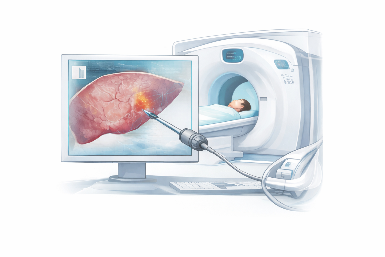
Imaging-guided tumour ablation is a modern, minimally invasive treatment used to destroy selected tumours using heat or cold, without the need for open surgery. At Sydney Interventional Radiology (SIR), this advanced treatment is performed by specialist interventional radiologists using real-time medical imaging for precision and safety.
Tumour ablation is commonly used for selected tumours in the liver, kidneys, and lungs. In appropriately selected patients, it can be as effective as conventional surgical removal.